
Specialist Technology For Precision Scleral & Corneal Lens Fitting
Specialist Technology For Precision Scleral & Corneal Lens Fitting
Specialist Technology For Precision Scleral & Corneal Lens Fitting
Specialist Technology For Precision Scleral & Corneal Lens Fitting
Assessing Vision
Patients often see streaks, doubling and haloes – even with their contact lenses. We have the technology to measure these optical distortions and objectively assess and modify the performance of the contact lens to provide better vision.
iTrace Ray-tracing
The world’s most advanced aberrometer maps the position of 256 low power lasers as they travel through the eye onto the retina in less than a second. The iTrace allows Dr. Dave to evaluate the distortions induced by the cornea and internal components of the eye.
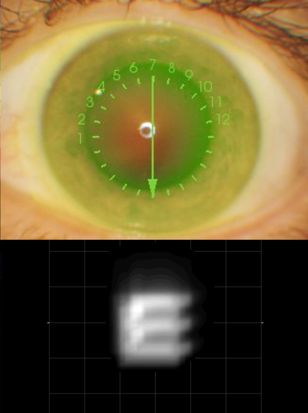
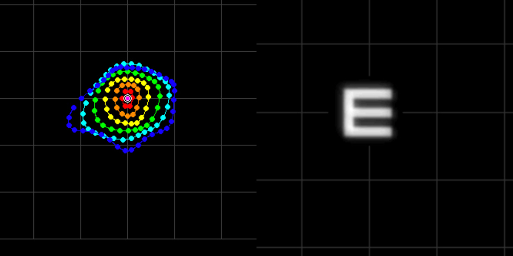
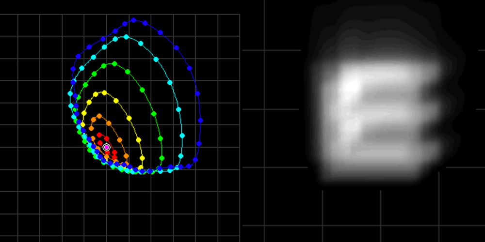
Improving Vision
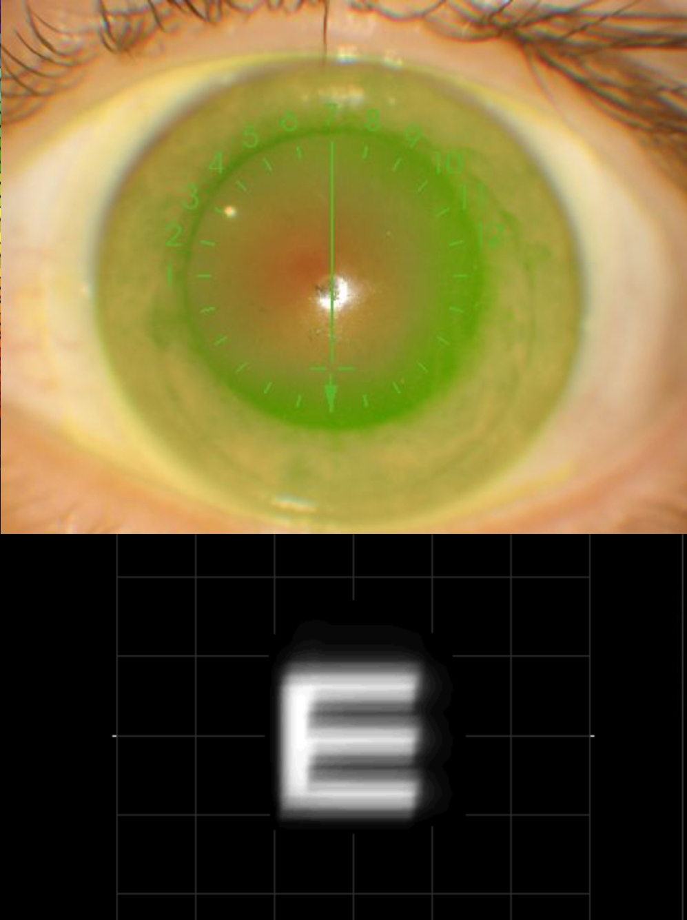
By performing aberrometry over a custom designed lens allows Dr. Dave to see how you see through the lens. Different curves on the front and back surface of the lens affect the size and intensity of visual distortions. By manipulating the fit, Dr. Dave can optimise vision.
Dr. Dave is able to precisely design freeform scleral and corneal lenses and additionally place optical corrections like astigmatism on the front or back surface of the lens.
Improving Vision
By performing aberrometry over a custom designed lens allows Dr. Dave to see how you see through the lens. Different curves on the front and back surface of the lens affect the size and intensity of visual distortions. By manipulating the fit, Dr. Dave can optimise vision.
Dr. Dave is able to precisely design freeform scleral and corneal lenses and additionally place optical corrections like astigmatism on the front or back surface of the lens.
Assessing Structure
The importance of assessing the structure of the cornea is critical for early diagnosis and to see which parts of the cornea are being affected.
Anterior OCT
OCT stands for Optical Coherence Tomography. This revolutionary technology allows Dr. Dave to identify the area on the cornea most affected by thinning or scarring.
Anterior OCT provides information within a structure, in this case the cornea.
Anterior OCT is also useful to evaluate the fitting of scleral lenses but it is not useful in designing the optimal fit of the lens to the eye.
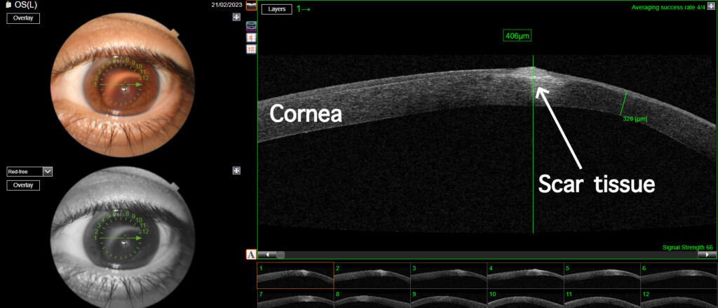
OCT scan showing thinning & scarring in Keratoconus
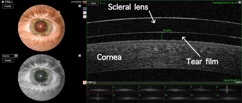
Scleral lens fitting can be accurately optimised
Specular Microscopy
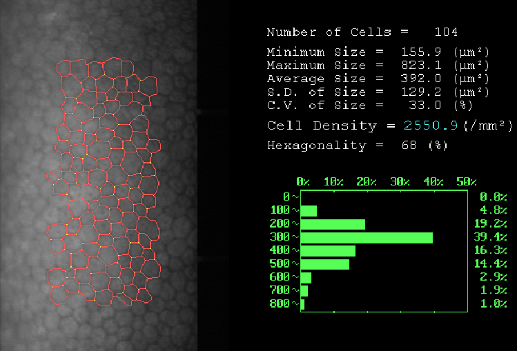
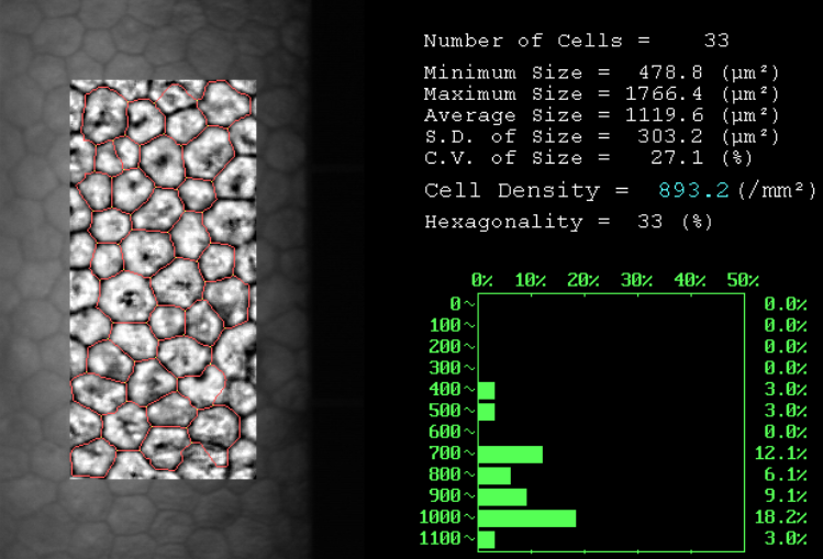
The cornea has special cells (endothelial cells) that constant pump water out to stop it from becoming cloudy.
As keraotconus progresses, the density of the cells decreases. Specular microscopy provides an automated endothelial cell count (ECC). We use this at initial visits to assess the health of the cornea.
The most important use of the ECC is when you have a corneal graft. A drop in ECC may indicate inflammation or even rejection that would indicate the need for steroid drops.
Specular Microscopy
The cornea has special cells (endothelial cells) that constant pump water out to stop it from becoming cloudy.
As keraotconus progresses, the density of the cells decreases. Specular microscopy provides an automated endothelial cell count (ECC). We use this at initial visits to assess the health of the cornea.
The most important use of the ECC is when you have a corneal graft. A drop in ECC may indicate inflammation or even rejection that would indicate the need for steroid drops.
Assessing Shape
Measuring the shape of the cornea and sclera is essential for accurate fitting of scleral and corneal lenses. Measuring corneal shape allows accurate monitoring of corneal disease.
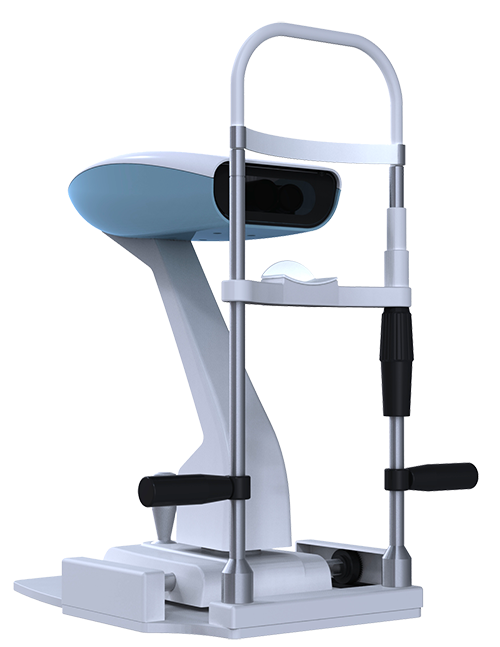
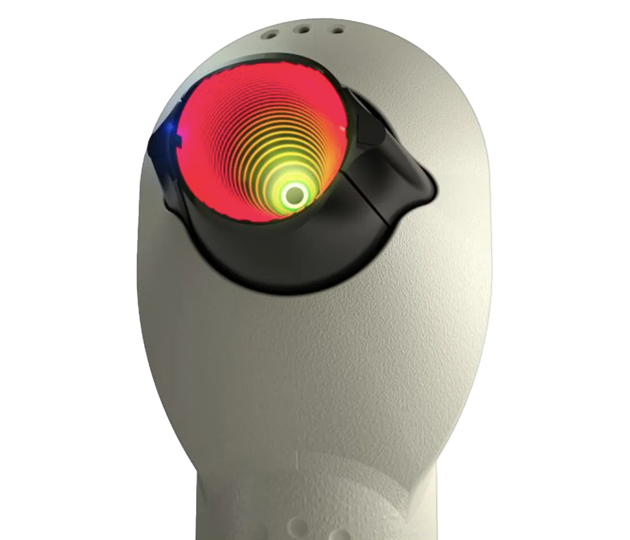
Profilometry
The only technology in the world that measures up to 20mm diameter of the front of the eye in a single capture. Over 350,000 points are captured to produce a precise 3D surface profile of your eye without touching the eye!
Why?
Scleral lenses rest entirely on the sclera (the white of the eye) so knowing the precise shape of the sclera allows us to make a lens to match the shape of the eye. When a lens doesn’t match the shape of the sclera, it often results in reduced vision, lens fogging and reduced wearing time. Until now, patients either have scleral lenses fitted by trial and error (the vast majority) or in some cases a cast of the eye is taken (expensive and also prone to error from contraction of the cast and eye movements causing errors in the cast).
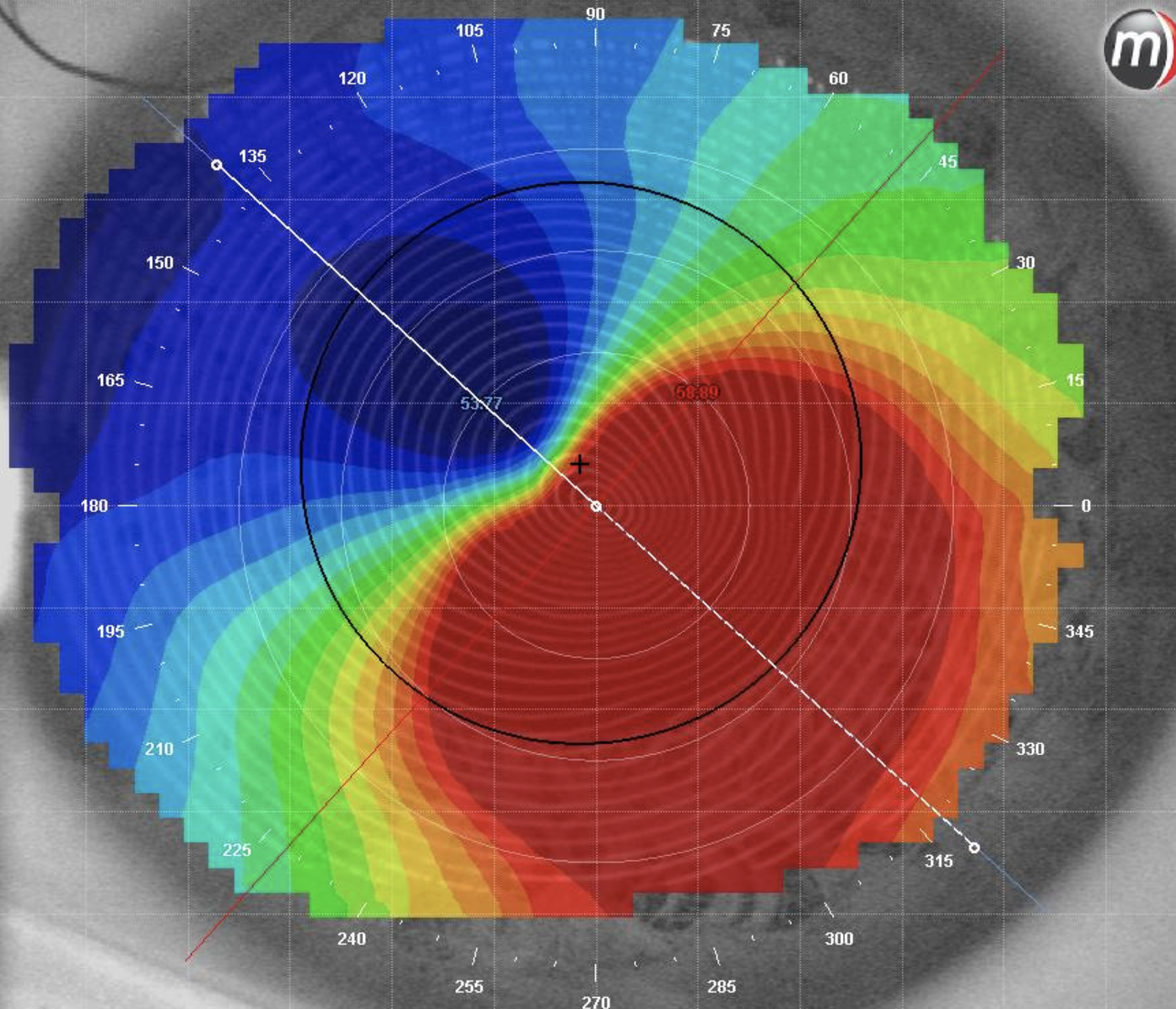
Corneal Topography
Subtle changes to the cornea are evaluated using both Profilometry and Corneal topography. Corneal topography assists in the design of rigid gas permeable lenses. We also use our freeform design process to make rigid gas permeable lenses that align precisely with the asymmetric cornea resulting better comfort. Corneal topography only measures the shape of the cornea and is not helpful in scleral lens fitting.

